Poster (Scientific congresses and symposiums)
Multiple Material Mesh Generation For Biomedical Applications
D'Otreppe, Vinciane; Boman, Romain; Ponthot, Jean-Philippe
2010National Committee on Biomedical Engineering - National Day
The finite element (FE) method is commonly used in biomedical applications for the simulation of the behaviour of biological structures. A key component in FE simulation is the creation of a finite element mesh. In medical applications, the meshes should be directly generated from the medical scans. Moreover, biological structures are usually composed of several inner regions that need to be separately segmented, labelled and meshed to be able to apply different material properties in the finite element model. A procedure to create surface meshes from a multi-valued volume data sets is proposed. Following properties are guaranteed: (1) The generated mesh consists of a set of non manifold triangle meshes that separate each connected component in the labelled data set. These interface meshes join each other consistently along their boundaries, i.e., no T junctions nor gaps may appear. (2) The surface mesh is a geometrically accurate representation of the data represented in the medical scans. However, it is not be tainted by the typical aliasing and staircase artifacts that are due to the discrete nature of the voxels.
Master’s dissertation (Dissertations and theses)
Génération de maillages surfaciques pour la création de modèles biomécaniques du cerveau
D'Otreppe, Vinciane
2008
Le planning préopératoire en neurochirurgie est réalisé à partir d'images structurelles et fonctionnelles du patient. Cependant l'intervention chirurgicale réalisée à partir de ce planning se fonde sur l'hypothèse que les structures anatomiques ne bougent pas pendant l'opération. En réalité, pendant l'opération, le cerveau se déforme de sorte que les images préopératoires, sur lesquelles se base le neurochirurgien, ne correspondent plus à la réalité anatomique du patient. Un approche envisagée pour résoudre ce problème est de modéliser, par la méthode des éléments finis, le comportement mécanique du cerveau pendant l'opération. Ceci permettrait de fournir au chirurgien, tout au long de l'intervention, des images mises à jour de la même qualité que les images préopératoires. La création du maillage du modèle biomécanique du cerveau à partir d'images IRM préopératoires fait l'objet de ce travail. La première et majeure partie du travail est consacrée à l'élaboration d'un mailleur surfacique. L'extraction de la surface, définie soit à l'aide d'une fonction implicite, soit à l'aide d'une image tridimensionnelle, se fait au moyen de la méthode Marching Tetrahedra. Le maillage ainsi obtenu étant inadapté au calcul éléments finis, il est ensuite simplifié et amélioré de manière à obtenir un maillage de bonne qualité. Afin de découpler la taille des mailles générées de la distance inter-slices, une méthode d'interpolation entre images a également été implémentée. La seconde partie est l'intégration du mailleur surfacique créé dans l'embryon du système de neuronavigation en cours de développement à l'ULg. Par simplicité, le cerveau est considéré comme un milieu homogène et seul le phénomène du brain shift est modélisé. De plus, les images IRM sont supposées avoir été préalablement corrigées, segmentées et recalées. Les résultats obtenus après calcul éléments finis similaires à ceux trouvés dans la littérature.
Doctoral thesis (Dissertations and theses)
From medical imaging to finite element simulations: a contribution to mesh generation and locking-free formulations for tetrahedra
D'Otreppe, Vinciane
2012
Patient-specific finite element (FE) modelling is gaining more and more attention over the years because of its potential to improve clinical treatment and surgical outcomes. Thanks to patient-specific modelling, the design of individualised implants and prostheses, surgical pre-operative planning and simulation, and the computation of stresses and strains in a patient's organ for diagnostic purposes will become a reality in the future. This work investigates two of the most challenging tasks of patient-specific modelling: the creation of image-based finite element meshes and the development of a low-order locking-free tetrahedral element. 
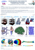
First, a general meshing strategy for tetrahedral mesh generation from segmented 3D images is proposed. The originality of the approach is the addition of surface reconstruction algorithm to the traditional image-to-mesh pipeline. The main advantages for this are: the generation of smooth boundaries, robustness to segmentation noise, a user-defined mesh resolution and a good fidelity of the mesh boundaries with respect to the underlying image. Also, the proposed meshing strategy is capable of generating meshes of heterogeneous structures, containing several interconnected types of tissues. Applications demonstrate that the interfaces between distinct material regions are topologically correct, i.e. the connections are edge-on-edge and node-on-node. Specific mesh decimation and mesh smoothing algorithms were designed for this multi-material tetrahedral mesh generator. In a last chapter, patient-specific hexahedral meshes are created by combining the proposed surface reconstruction algorithm with a classical voxel-conversion algorithm. Second, a low-order tetrahedral element for the solution of solid mechanics problems involving nearly incompressible materials is developed. The formulation is based on F-bar methodologies and nodal-based formulations. As in nodal based formulations, nodal Jacobians are defined. These nodal quantities are then averaged over the element to define a modified elemental Jacobian, which is used to define a modified deformation gradient, F-bar, for the element. Both 2D triangular and 3D tetrahedral are proposed and they can be used for both implicit and explicit analysis. The exact stiffness terms for the tangent stiffness matrix are derived so that a quadratic convergence rate in ensured for the Newton-Raphson equilibrium iterations. Most importantly, the new element can be used regardless the material model. Benchmarking 2D and 3D numerical tests using several constitutive models indicate a substantial removing of both the volumetric and the shear locking tendency of the standard linear triangle and tetrahedron, as well as an accurate distribution of strain, stress and pressure fields. The potential of the resulting image - to - FE model procedure is demonstrated in the last part of this work, through patient-specific finite element analyses of actual biomechanical research topics.
Paper published in a book (Scientific congresses and symposiums)
Development of a biomechanical model of deer antler cancellous bone based on X-ray microtomographic images
de Bien, Charlotte; Mengoni, Marlène; D'Otreppe, Vinciane et al.
2012In Micro-CT User Meeting 2012 - Abstract Book
Finite element models accurately compute the mechanical response of bone and bone-like materials when the models include their detailed microstructure. The aim of this paper is to develop and validate a biomechanical model for deer antler cancellous bone tissue based on X-ray microtomographic images. In order to simulate the mechanical behavior under compressive load using a finite element model, images obtained by X-ray microtomography were exported into Metafor, which is an non-linear finite element software initially developed at University of Liège for metal forming processes. This software has recently found biomedical applications. The ultimate goal is to compare model predictions with the mechanical behavior observed experimentally using the Skyscan material testing stage under compression mode. The creation of the biomechanical model mesh from segmented μCT images, its integration into the software Metafor and the simulation of a compression test are described in this paper.
Peer reviewed
Unpublished conference/Abstract (Scientific congresses and symposiums)
Finite element simulation of canine humeral condylar fractures
D'Otreppe, Vinciane; Böhme, Béatrice; Balligand, Marc et al.
2011ISB 2011
Humeral condylar fractures are common in dogs. Different types of fractures (lateral, medial, bicondylar) may occur, depending on the age of the dog and the position of its elbow during the impact. The goal of this work is to understand the effects of bone posture and skeletal development on canine humeral fractures by means of the finite element method. Four distinct finite element simulations were performed, corresponding to an immature and a mature dog elbow, respectively in extension and flexion. To create the finite element models, subject-specific finite element meshes were extracted from the CT-data. Appropriate material properties were used for cortical bone, trabecular bone and cartilage. The modified Mohr-Coulomb failure criterion was implemented to take account for strength asymmetry. Lateral humeral fractures are obtained for both the young and adult dog elbow, in extension and flexion. This is in agreement with clinical observations, in which lateral condylar fractures are most common.
Paper published in a book (Scientific congresses and symposiums)
Génération de maillages pour la creation de modèles biomécaniques du cerveau
D'Otreppe, Vinciane; Boman, Romain; Ponthot, Jean-Philippe
2009In Raous, Michel; Pasquet, Philippe; Rey, Christian (Eds.) Neuvième Colloque National en Calcul des Structures
Nous présentons une méthode efficace et automatique pour générer des maillages éléments finis surfaciques de haute qualité et de bonne fidélité géométrique au départ d’images médicales segmentées. Cette méthode est basée sur l’algorithme du Marching Tetrahedra auquel nous avons adjoint des techniques permettant d’ajuster la topologie et les positions nodales du maillage afin de renforcer la qualité des éléments tout en respectant la géométrie de l’objet présent dans les images. Le but ultime de l’approche est la modélisation du cerveau lors d’interventions neurochirurgicales.
Poster (Scientific congresses and symposiums)
Génération de Maillages pour la Création de Modèles Biomécanique du Cerveau
D'Otreppe, Vinciane
20099ième Colloque National en Calcul des Structures
Unpublished conference/Abstract (Scientific congresses and symposiums)
Image-based Finite Element Mesh Generation for Microstructures
D'Otreppe, Vinciane; Mengoni, Marlène; Boman, Romain et al.
2011Fifth International Conference on Advanced COmputational Methods in ENgineering (ACOMEN 2011)
Paper published in a book (Scientific congresses and symposiums)
Smooth multiple-region mesh generation for biomedical applications
D'Otreppe, Vinciane; Boman, Romain; Ponthot, Jean-Philippe
2010In ECCM 2010, 4th European Conference on Computational Mechanics
The finite element method is commonly used in biomedical applications for the simulation of the behaviour of biological structures. However, extracting finite element meshes from medical data is still very challenging. In this article, an innovative system that efficiently reconstructs smooth, multi-material, 3D surface meshes from medical images is presented. Our approach is based on an enhanced Marching Tetrahedra algorithm, which extracts boundary surfaces between different materials within one sweep of the image stack in an integrated manner. Moreover,Multi-Level Partition of Unity implicit models are used to obtain a smooth and accurate surface representation of the original binary sampled surface. Mesh smoothing and decimation algorithms were also revised to adapt to the multi-material nature of the system as well as to adhere to the underlying volume data.
Peer reviewed
Paper published in a book (Scientific congresses and symposiums)
Multiple material mesh generation for biomedical applications
D'Otreppe, Vinciane; Boman, Romain; Ponthot, Jean-Philippe
2010In Proceedings of the 9th belgian day on biomedical engineering
The finite element method is commonly used in biomedical applications for the simulation of the behaviour of biological structures. However, extracting finite element meshes from medical images is still very challenging. We propose an innovative system to create accurate multiple domain tetrahedral meshes from medical images. Our approach is based on an enhanced Marching Tetrahedra algorithm that extracts the boundary surfaces delimiting the different material domains in an integrated manner. Moreover, a surface reconstruction method is employed to ensure that the resulting mesh is a smooth and accurate surface representation of the original sampled structure. Mesh smoothing and decimation algorithms are also revised to conform to the multiple material nature of the system as well as to adhere to the underlying volume data.
Peer reviewed
Paper published in a book (Scientific congresses and symposiums)
A fully nonlinear finite element model for orthodontic tooth movement prediction
Mengoni, Marlène; D'Otreppe, Vinciane; Ponthot, Jean-Philippe
2012In Proceedings of the 10th International Symposium on Computer Methods in Biomechanics and Biomedical Engineering.
Peer reviewed
Unpublished conference/Abstract (Scientific congresses and symposiums)
On the periodontal ligament representation in 3D finite element analysis for orthodontic tooth movement models
Mengoni, Marlène; D'Otreppe, Vinciane; Ponthot, Jean-Philippe
2011Fifth International Conference on Advanced COmputational Methods in ENgineering (ACOMEN 2011)
Paper published in a book (Scientific congresses and symposiums)
Generating smooth surface meshes from labelled medical datasets
D'Otreppe, Vinciane; Boman, Romain; Ponthot, Jean-Philippe
2011In Nithiarasu, Perumal; Löhner, Rainald; van Loon, Raoul et al. (Eds.) CMBE11: 2nd International Conference on Computational & Mathematical Biomedical Engineering, 30 March - 1 April, 2011, Washington D.C. U.S.A.
In this article, an innovative procedure to create high quality triangular meshes from medical scans is proposed. The approach is based on an enhanced Marching Tetrahedra algorithm that extracts a consistent multiple region surface mesh from a labelled volume data set; coupled with a surface reconstruction method to avoid typical staircase artifacts. Mesh smoothing and decimation algorithms are also revised to conform to the multiple material nature of the system as well as to adhere to the underlying volume data. The proposed method is well suited for subsequent volume mesh generation and finite element simulations.
Peer reviewed
Unpublished conference/Abstract (Scientific congresses and symposiums)
Development of a thorax CT-scan based finite element model for thoracic injury assessment
Nsiampa, Nestor; D'Otreppe, Vinciane; Robbe, Cyril et al.
2011Advanced COmputational Methods in ENgineering (ACOMEN 2011)
Poster (Scientific congresses and symposiums)
Considerations on the Pathophysiology of Canine Condylar Fractures by Finite Element Analysis
Böhme, Béatrice; D'Otreppe, Vinciane; Ponthot, Jean-Philippe et al.
201120th Annual Scientific Meeting of the European College of Veterinary Surgeons
Paper published in a journal (Scientific congresses and symposiums)
A bone remodelling model for long term orthodontic tooth movement.
Mengoni, Marlène; D'Otreppe, Vinciane; Ponthot, Jean-Philippe
2012In Journal of Biomechanics, 45 (Supplement 1), p. 180
Peer Reviewed verified by ORBi
Article (Scientific journals)
Generating smooth surface meshes from multi-region medical images
D'Otreppe, Vinciane; Boman, Romain; Ponthot, Jean-Philippe
2012In International Journal for Numerical Methods in Biomedical Engineering, 28 (6-7), p. 642-660
Thanks to advances in medical imaging technologies and numerical methods, Patient-Specific Modelling is more and more used to improve diagnosis and to estimate the outcome of surgical interventions. It requires the extraction of the domain of interest from the medical scans of the patient, as well as the discretisation of this geometry. However, extracting smooth multi-material meshes that conform to the tissue boundaries described in the segmented image is still an active field of research. We propose to solve this issue by combining an implicit surface reconstruction method with a multi-region mesh extraction scheme. The surface reconstruction algorithm is based on multi-level partition of unity implicit surfaces which we extended to the multi-material case. The mesh generation algorithm consists in a novel multidomain version of the marching tetrahedra. It generates multi-region meshes as a set of triangular surface patches consistently joining each other at material junctions. This paper presents this original meshing strategy, starting from boundary points extraction from the segmented data, to heterogeneous implicit surface definition, multi-region surface triangulation and mesh adaptation. Results indicate that the proposed approach produces smooth and high-quality triangular meshes with a reasonable geometric accuracy. Hence, the proposed method is well suited for subsequent volume mesh generation and Finite Element simulations.
Peer Reviewed verified by ORBi