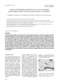
[en] Ultrasound has become more common in the investigation of the podotrochlear apparatus in the horse and the use of more sensible scanheads improved
the quality of the images. This study shows six examples of the ultrasound anatomy of the distal
podotrochlear apparatus in the transcuneal view performed with three different scanheads. The
presented transcuneal scans of the podotrochlear apparatus of an 18 months old warmblood
foal have been established post mortem on isolated feet. Modus of registration was real-timemotion, linear- and sectorscanheads working at 7.5 (linear)
and 7.5 (microconvex) and 3.5 (convex) MHz were used. The echoanatomic structures of the
soft tissue and bony surfaces in the distal podotrochlear region are compared by six images.
The combination of the different scanheads permits an improved complete visualisation in this
area. Therefore these ultrasound pictures show that ultrasonography is an useful complementary
investigation method that enables to precise the diagnosis
Research center :
Centre Européen du Cheval de Mont-le-Soie, 1 Mont-le-Soie, 6690 Vielsalm, Belgique Service d’Imagerie médicale, Faculté de Médecine vétérinaire, Université de Liège, 20 Boulevard de Colonster, 4000 Liège, Belgique Secteur d'Anesthésiologie générale et Pathologie chirurgicale, Pôle équin, Clinique des animaux de compagnie et des équidés, Faculté de Médecine vétérinaire, Université de Liège, 20 Boulevard de Colonster, 4000 Liège, Belgique Unité mixte de Recherche INRA-ENVA, Laboratoire de Biomécanique et Pathologie locomotrice du Cheval, Clinique équine, CIRALE, Ecole nationale vétérinaire d’Alfort, 7 Avenue du Général de Gaulle, 94704 Maisons-Alfort, France Institut für Veterinäranatomie, 20 Koserstrasse, FU-Berlin, 14195 Berlin, Germany
Disciplines :
Veterinary medicine & animal health
Author, co-author :
Schneider, Nicole ; Université de Liège - ULiège > CARE "Le Réseau des bibliothèques" > Bibliothèque des Sciences de la vie
Busoni, Valeria ; Université de Liège - ULiège > Département clinique des animaux de compagnie et des équidés > Imagerie médicale
de la Rebière de Pouyade, Geoffroy ; Université de Liège - ULiège > Département clinique des animaux de compagnie et des équidés > Anesthésiologie gén. et pathologie chirurg. des grds animaux
Denoix, Jean-Marie
Weyrauch, K. D.
Language :
French
Title :
Examen échographique transcunéen post mortem de l'appareil podotrochléaire équin à l'aide des sondes linéaires et sectorielles
Alternative titles :
[en] Post mortem transcuneal ultrasound study of the equine podotrochlear apparatus by linear and sectorial scanheads.
Blunden A., Dyson S., Murray R., Schramme M. Histopathology in horses with chronic palmar foot pain and age-matched controls. Part 1: navicular bone and related structures. Equine Vet. J., 2006, 38, 15-22.
Bolen G., Busoni V., Jacqmot O., Snaps F. Sonographic anatomy of the palmarodistal aspect of the equine digit. Vet. Radiol. Ultrasound, 2007, 48, 270-275.
Busoni V., Denoix J.M. Diagnostic imaging of the equine foot. In: 6° Congresso Nazionale Multisala Sive, Pisa, 29-31st Gennaio 2000, 31-40.
Busoni V., Denoix J.M. Ultrasonography of the podotrochlear apparatus in the horse using a transcuneal approach: technique and reference images. Vet. Radiol. Ultrasound, 2001, 42, 534-540.
Busoni V., Mean MN., Brigone L., Snaps F. Echographie de l'appareil podotrochléaire: étude in vitro sur 30 membres isolés de cheval. Ann. Méd. Vét., 2002, 146, 181-187.
Busoni V., Heimann M., Trenteseaux J., Snaps F., Dondelinger R.F. Magnetic resonance imaging findings in the equine deep digital flexor tendon and distal sesamoid bone in advanced navicular disease: an ex vivo study. Vet. Radiol. Ultrasound, 2005, 46, 279-286.
Caudron I. Approche orthopédique des affections ostéoarticulaires dégénératives de l'extrémité digitale du cheval: prévention et traitement. (PhD thesis). Université de Liège: Liège, 1998, 229 p.
Denoix J.M., Perrot P., Bousseau B. Crevier N. Apport de l'échographie dans le diagnostic des affections articulaires chez le cheval. Point Vét., 1994, 26, 197-206.
Denoix J.M., Tappert J., Mathieu D. Magnetic resonance imaging of the foot: will this revolutionise diagnosis? In: 38th British Equine Veterinary Association (BEVA) Congress, 12-15th September 1999, 44-45.
Dyson S., Murray R., Schramme M., Branch M. Lameness in 46 horses associcated with deep digital flexor tendonitis in the digit: diagnosis confirmed with magnetic resonance imaging. Equine Vet. J., 2003, 35, 681-690.
Jackmann B.R., Baxter G.M., Doran R.E., Douglas A., Parks A.H. Palmar digital neurectomy in horses. Vet. Surg., 22, 1993, 285-288.
Keller H., Grundmann S. Radiologische Ver lauf sunter suchung der Podotrochlose. Tierärztl. Prax., 1995, 23, 46-52.
Kotani H., Taura Y., Sakai A., Tsuka T., Kageyama Y. Nakaichi M. Antemortem evaluation for magnetic resonance of the equine flexor tendon. J. Vet. Med. Sci., 2000, 62, 81-84.
Murray R.C., Bridget L., Roberts M.C. Schramme M., Dyson S., Branch M. Quantitative evaluation of equine deep digital flexor tendon morphology using magnetic resonance imaging. Vet. Radiol. Ultrasound, 2004, 45, 103-111.
Poulos P.W., Brown A., Brown E., Gamboa L. On navicular disease of the horse. Vet. Radiol., 1998, 30, 54-58.
Sage A.M., Turner T.A. Ultrasonography of the soft tissue structures of the equine foot. Equine Vet. Educ., 2002, 14, 221-224.
Schneider N. Studie zur sonographischen Anatomie des Hufgelenks und der Hufrolle beim Pferd-ein Beitrag zur Diagnose der Podotrochlose (Inaugural Dissertation). Freie Universität Berlin: Berlin, 2003, 167 p.
Serteyn D., Vanschaepdael P., Caudron I., Miesen M., Lalet P., Grulke S., Staquez S. Quelques notions de l'extérieur du cheval: évaluation clinique de la ferrure Equi + lors de pathologies de l'articulation interphalangienne distale. Prat. Vét. Equine, 1995, 27, 105-110.