Abstract :
[en] Nowadays “Unified Segmentation” (US) is the usual approach to warp brain images into a standard reference space, i.e. perform spatial normalization, and derive posterior probability maps of the brain tissues, typically grey and white matter (GM, WM) and CSF [1]. US only relies on a spatial deformation model and prior ‘tissue probability maps’ (TPM) of the head tissues.
When dealing with data from patients with focal brain lesions, e.g. tumors or multiple sclerosis (MS) lesion, the standard US approach does not work as it cannot account for the abnormal tissue distribution. A common work around is “cost function masking” (CFM) [2,3] where the abnormal tissues are masked out using a binary mask of the lesion [6,8].
Here we extend the US approach to provide a more principled solution for brain images with focal lesions. The aim is twofold: a more accurate warping into the reference space of the healthy tissues and a more precise delineation of the lesion(s).
We modify the standard TPM adding a subject-specific ‘lesion probability map’ [5,7], by
1/ estimating a preliminary spatial warping from subject to the reference space with the CFM approach, then 2/ carefully updating the TPM with a new tissue class, the lesion, defined from the smoothed warped lesion mask and deciding which healthy tissue class can be affected by the lesion.
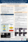
The TPM-with-lesion is then fed into the US with the patients images, see Fig. 1. This “US-with-Lesion” (USwL) approach thus accounts for the presence of focal abnormal tissues in a probabilistic way, providing posterior probability maps of the tissues, including the lesion, and spatial deformation, accounting for the lesion. We tested and evaluated our USwL approach on 2 publicly available datasets: the BRATS [4] and the ‘MS lesion segmentation challenge’ (MSchal)[8]. The BRATS data include T1 and FLAIR images of 30 patients with gliomas and their annotated tumor mask (further considered as the ground truth). A rough lesion mask was manually built from the FLAIR image using MRIcron. USwL was used to segment T1 and FLAIR images along with this approximate mask. The GM, WM and CSF tissue classes could be affected by the lesion. The posterior probability map for the lesion tissue was cleaned up (preserving the bigger clusters) and thresholded. Overall the USwL improved (p<.05) the similarity of the lesion mask to the annotated tumor, in term of voxel matching (sensitivity, specificity & Jaccard coefficient). Synthetic lesioned brains were also generated to assess the quality of the deformation for the healthy tissues, indicating the superiority (p<.05) of the USwL compared to the standard approach.
The MSchal data include T1, T2 and FLAIR images of 20 patients with MS as well as the manually annotated lesion (considered as only approximate here). USwL is applied on the 3 structural images with the lesion mask provided and with the constraint that only the WM is potentially affected by the lesion (as is plausible with MS). The thresholded posterior probability map for the lesion tissue was compared to the provided lesion mask. The USwL lead to more biologically plausible lesion volumes (p<.05), in term of volume compactness [10], see Fig. 2. The similarity of the warped posterior GM maps across the 20 subjects (expressed as the root-mean square difference to the mean of the 20 subjects)
was also examined. The improvement, from using CFM-US to USwL, in the between-subject GM-matching is proportional (p<.05) to the actual WM lesion volume.
We provide a new tool for US that allows to include focal lesions. Over the 2 dataset considered, USwL demonstrated improved performances compared to the standard US: 1/ a more accurate warping into the reference space of the healthy tissues and 2/ simply using an approximate mask, a more precise delineation of the lesion(s). The whole code will be made available as an SPM add-on toolbox (with a batch interface) on
https://github.com/CyclotronResearchCentre/USwLesion.