Abstract :
[en] Introduction
A single tumoral tissue can bear phenotypically different cell populations. This phenomenon called intra-tumor heterogeneity can lead to differential behaviors regarding metastasis seeding and therapy resistance [Zardavas et al., Nature Rev. Clin. Onc. 2015]. MALDI imaging has proven its efficiency for revealing hidden molecular features offering an insight into distinct cellular regions based on their molecular content. Further, proteomics applied to these regions could allow depicting the molecular context associated to particular cells groups and enable the collection of qualitative, quantitative and spatial information for each protein.
Methods
Breast cancer Formalin Fixed and Paraffin Embedded tissues, from patients whose outcome had been recorded over a period of 10 years, were provided by the department of Pathology of University of Liège. After Citric Acid Antigen Retrieval and trypsin digestion, images were obtained by MALDI-TOF/TOF-MS (Bruker, Germany). From the obtained datasets, segmentation and analytical data analysis were applied using SCiLS (Bruker, Germany) and the cloud software Multimaging (ImaBiotech, France).
Small tissue areas were obtained by laser microdissection (LEICA LMD 700, Germany), upon which a combination of chemical processes was applied to ensure optimal protein antigen retrieval, extraction and digestion. Finally, the tissue pieces obtained were analyzed by LC-MS/MS using UPLC Waters Nanoacquity and Thermo Q-Exactive instruments.
Preliminary data
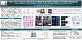
Based on mathematical calculations for the MALDI imaging datasets of the breast cancer FFPE tissues, Regions Of Interest (ROIs) were detected in a single tumor, revealing intra-tumoral heterogeneity, which can be correlated to the level of aggressiveness of the affliction and to the final prognosis of the patient. We aimed to compare the proteomic profiles of each of the small ROIs. Until today, proteomics applied to tissues composed by a restricted number of cells is quite tedious due to possible tissue losses during their handling. Recently, Longuespée [Longuespée et al., Methods 2015] published a method in order to retrieve the identification of 1400 proteins from microdissected tissue pieces containing only 2700 cells.
This whole procedure allowed us to identify a panel of protein that characterizes tissue heterogeneity within a single tumor. This proves the applicability of the combination of MALDI imaging for the discovery of intra-tumoral heterogeneity without a priori, on a mathematical basis, and classical proteomics applied on laser-microdissected tissue samples of very restricted areas. This method will now be applied to several MALDI datasets in order to retrieve commune ROIs and to associate their presence with the information of each patient, such as their prognosis. Those ROIs will then be microdissected and subjected to microproteomic methods that will allow us to retrieve the extensive molecular context associated to bad patient prognosis and/or therapy resistance. The possibility to identify protein/peptide markers will have the power to predict the outcome of the breast cancer patient at the beginning of their treatment, and thus, improve the clinical care for the benefit of the patients.
Novel aspect
The workflow combines the unique advantages of MALDI imaging for de novo molecular features characterization and LMD-based microproteomics.
Disciplines :
Physical, chemical, mathematical & earth Sciences: Multidisciplinary, general & others