[en] The supporting cells of the hearing organ are characterized by the presence of an abundant cytoskeleton which is mainly composed of microtubules. These supporting cells have also been shown to contain a minor mammalian tubulin, the β5-tubulin, recently reported as a biomarker of cell proliferation. It was shown that a β-tubulin isoform can specify the microtubule architecture, as seen with the expression of the Moth β2 tubulin in the Drosophila testes which imposes the 16-protofilament (16pf) structure on the corresponding subset of Drosophila microtubules. Moreover, supporting cell microtubules are formed by 15pf instead of the canonical 13, a unique fact among vertebrates. Such a protofilament configuration has been observed in C. elegans’ neurons which are responsible for the mechanosensory sense of touch. It was also shown that these 15pf microtubules were essential to the proper functioning of these neurons.
To determine the role of this particular tubulin in the auditory organ and its possible involvement in the formation of the unusual 15pf microtubules of supporting cells, we studied the spatiotemporal localization of β5-tubulin during development in rats from embryonic day 18 until P25 (25th postnatal day). Then we examined the fine structure of microtubules at the transmission electron microscope level (TEM).
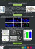
Our results showed that β5-tubulin, contrary to other β-tubulins, had a unique distribution in the cochlea. This β-tubulin appeared at a postnatal stage, before the opening of the Corti’s tunnel and is restricted to supporting cells, especially in pillar and Deiters’ cells. Our TEM study further indicated that these cells were composed by 13pf microtubules at P2, but by 15pf microtubules at P25.
In conclusion, the architecture and composition of microtubules present in the supporting cells change during development of the Corti organ.
Further experiments are now required to determine if these changes are related to the appearance of β5-tubulin.