[en] PET studies provide valuable information in the assessment of animal models for human diseases.
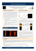
MicroPET systems provide the high resolution needed to explore small organs but suffer from a reduced axial FOV. Multiple bed positions are then used to obtain whole body scans resulting in increased scan time and incomplete dynamic data. In contrast, human PET systems have larger axial FOV but a lower resolution. In this study, an image-based model of the scanner spatial response function combined with a 3D-OSEM reconstruction algorithm were used to improve spatial resolution of the Siemens ECAT EXACT HR+ PET scanner. A stationary double Gaussian model [1] of the ECAT EXACT HR+ point spread function was derived from 18F point source measurements performed at different radial and axial locations in the scanner FOV. This model was used in a 3D-OSEM reconstruction (3D-OSEM-RM). Sinograms were normalized and attenuation and scatter corrected using the Siemens ECAT tools before reconstruction. Both NEMA NU 2-1994 performance phantoms and NEMA NU4-2008 image quality phantom mimicking small animals were used to evaluate the accuracy of corrections for physical effects and the overall image quality. A 50 min dynamic FDG rat study was conducted on the ECAT HR+ and reconstructed with 3D-OSEM-RM. The images were used to compute the metabolic rate of glucose (MRglu) in multiple brain structures. These images were also visually compared to the static image obtained with a FOCUS 120 microPET immediately after the HR+ dynamic scan. The standard deviations of the two Gaussians used to model the transaxial (axial) resolution in a central FOV of 5 cm radius were σ1 = 1.6 (2.75) mm and σ2 = 3.66 (4.16) mm, and the ratio of the weights between the first and second Gaussians was ρ = 0.2 (0.7). Image uniformity and accuracy of scatter and attenuation corrections, evaluated following NEMA NU 2-1994, were found to be very similar between 3D-OSEM, 3D-OSEM-RM, 2D- and 3D-FBP reconstructed images. When using the NEMA NU4-2008 image quality phantom a significant increase of the hot rod recovery coefficient was observed. This effect was rod size dependent and amounted to 17-35% for the 3D-OSEM-RM compared to the 3D-OSEM and to 35-62% compared to the FBP reconstructions. Nevertheless the values obtained with 3D-OSEM-RM were around 20-35% lower than those obtained with the FOCUS 120 microPET scanner. Most of the small brain structures observed on microPET images were also visible on the images obtained with the HR+ scanner and 3D-OSEM-RM. Rat cerebral MRglu values calculated on 3D-OSEM-RM images were in the range of published values [2] (e.g. whole brain = 25.34 μmol/min/100g). Using an approximate model of the ECAT EXACT HR+ spatial response in 3D-OSEM resulted in sufficient image quality for dynamic whole body scans of small rodents, despite the large FOV, and resulted in improved contrast compared to images generated using the built-in software. This methodology will be applied for future small animal dosimetry and modeling studies in our laboratory. [1] Comtat et al. IEEE Nucl Sci Symp Conf Record. pp. 4120-4123 (2008) [2] Schiffer et al. J Nucl Med 48:277-287 (2007)
Research center :
GIGA CRC (Cyclotron Research Center) In vivo Imaging-Aging & Memory - ULiège
Disciplines :
Life sciences: Multidisciplinary, general & others
Author, co-author :
Bahri, Mohamed Ali ; Université de Liège - ULiège > Centre de recherches du cyclotron
Tombuloglu, S
Warnock, Geoffrey ; Université de Liège - ULiège > Centre de recherches du cyclotron
Taleb, Dounia ; Université de Liège - ULiège > Département de physique > Imagerie médicale expérimentale
Bretin, Florian ; Université de Liège - ULiège > Centre de recherches du cyclotron
Degueldre, Christian ; Université de Liège - ULiège > Centre de recherches du cyclotron
Luxen, André ; Université de Liège - ULiège > Département de chimie (sciences) > Chimie organique de synthèse
Salmon, Eric ; Université de Liège - ULiège > Département des sciences cliniques > Neuroimagerie des troubles de la mémoire et révalid. cogn.
Plenevaux, Alain ; Université de Liège - ULiège > Centre de recherches du cyclotron
Comtat, C
Seret, Alain ; Université de Liège - ULiège > Département de physique > Imagerie médicale expérimentale
Language :
English
Title :
Small animal imaging with human PET
Publication date :
September 2012
Number of pages :
A0
Event name :
World Molecular Imaging Congress
Event organizer :
World Molecular Imaging Society
Event place :
Dublin, Ireland
Event date :
04/09/2012 - 08/09/2012
Audience :
International
Funders :
F.R.S.-FNRS - Fonds de la Recherche Scientifique [BE]